Dear Scholars, We wish to inform you that Aster Publications is now Asterium Scientific Publications. This change reflects our ongoing growth and commitment to excellence. We appreciate your continued support and look forward to serving you as Asterium Scientific Publications.

Open Access

Metallic line in Chest Radiograph: Artifact

B Jayakrishnan, FRCP 1* , Boris Itkin, MD 2 Anitha Jose, BSc3, Sami M Bennji, FCP(SA)4

1Senior Consultant, Division of Pulmonology, Head & Neck and Thoracic Program, Sultan Qaboos Comprehensive Cancer Care and Research Centre, University Medical City, Muscat, Oman
2Senior Consultant Oncologist, Rare Tumours Program, Sultan Qaboos Comprehensive Cancer Care and Research Centre, University Medical City, Muscat, Oman
3Staff Nurse, Sultan Qaboos Comprehensive Cancer Care and Research Centre, University Medical City, Muscat, Oman
4Consultant, Division of Pulmonology, Head & Neck and Thoracic Program, Sultan Qaboos Comprehensive Cancer Care and Research Centre, University Medical City, Muscat, Oman

Keywords
: Artifact, Chest X-ray

BACKGROUND


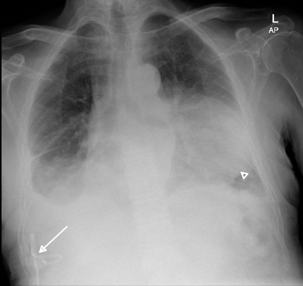
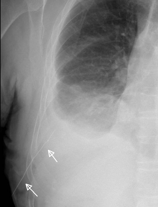
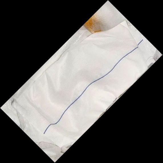
A 72-year-old man with advanced right thigh synovial sarcoma and pulmonary metastasis presented with bilateral pleural effusion in March 2024. Upon admission he underwent left-sided pleural drainage. A few days later, an 8F pigtail catheter was inserted on the right side, which successfully drained approximately 1500 ml of slightly blood-stained pleural fluid (Figure 1). However, on the same night, the catheter was inadvertently dislodged by the patient. A tight-fitting plaster was promptly applied, and he remained symptom-free. A follow up chest radiograph showed a clear linear radiopaque shadow measuring around 9.4 cm with the tip visible well inside the right chest (Figure 2). This shadow was puzzling as the pleural drain had been completely removed. There was no palpable swelling, wires, or tubes under the skin. Upon closer examination, it was discovered that the gauze used (Bromed Gauze Lap sponges, Brosco International Inc, Mesquite, USA) had a linear radiopaque marker (Figure 3). A repeat radiograph did not show this opacity. This radiopaque marker on the gauze resembled a metallic foreign object in the thorax, causing anxiety and confusion among the medical staff and the patient. Patient remained well without worsening of the effusion for two months but succumbed to his illness subsequently.

Figure 1: Chest radiograph showing a pigtail catheter on the right side (arrow), bilateral pleural effusions and a metastatic mass left lower zone (arrowhead).

Figure 1: Chest radiograph showing a pigtail catheter on the right side (arrow), bilateral pleural effusions and a metastatic mass left lower zone (arrowhead).


Figure 2: Right hemithorax view of the chest radiograph showing a white, radiopaque, straight line 9.4 cm suggestive of a thin wire on the right lower chest (arrows).


Figure 3: Sterile, super absorbent surgical sponge with a blue radiopaque strip.

DISCUSSION

An artifact on an image is a feature that does not correlate with the physical properties of the subject being imaged and may hinder an accurate diagnosis. Radiographic artifacts can result from hardware failure, operator errors or post-processing inaccuracies [1]. Common artefacts include motion distortion, radiopaque objects on/external to the patient, twin/double exposure, grid cut-off, debris in the housing and parallax effect. Notably, several artifacts on chest radiography have been reported to resemble pulmonary diseases. Skin folds, hair, clothing, and bed sheets can sometimes imitate pneumothorax; similarly, electrocardiographic leads and beads on clothing can mimic lung nodules [2-4]. In our patient the slant at which the artifact appeared coincided with the entry angle typically seen during pleural space access. Moreover, this specific gauze is not usually kept in our medical unit’s dressing sets and so the image was convincing for a procedural error. Often these artifacts can be deceptive, potentially leading to erroneous diagnoses and compromised patient care. In cases of uncertainty, physicians should exercise caution when planning interventions.


REFERENCES

  1. Shetty A, Torri G, Niknejad M, et al. X-ray artifacts. Reference article, Radiopaedia.org (Accessed on 18 Feb 2025).
  2. Jayakrishnan B, Al-Jamali A, Al-Mubaihsi S, Al-Aghbari J. Pneumothorax or Skin Fold? Mind the Gap. Sultan Qaboos Univ Med J. 2022; 22(4): 591–592.
  3. Kim T, Messersmith RN, MacMahon H. Pulmonary nodule mimicked by ECG lead artifact. Chest. 1989;95:237–238.
  4. Yoshihara S, Komagamine J. Clothing artefacts appearing as multiple lung pseudonodules. BMJ Case Rep. 2023;16: e257820.

Journal Name: Annals of Case Reports and Clinical Images

Article Type: Clinical  Image

Received: February 18, 2025; Accepted: February 19, 2025; Published: February 21, 2025

CC: This article is distributed under the terms of the Creative Commons Attribution License, which permits unrestricted use and redistribution provided that the original author and source are credited.

Competing interests: The authors have declared that no competing interests exist.

Cite as: Jayakrishnan B, Itkin B, Jose A, Bennji SM. Metallic line in Chest Radiograph: Artifact. Ann Case Rep Clin Images. 2025; 1:1-3.

Dr. B. Jayakrishnan
MBBS, MD,DTCD,DNB,MRCP(UK),FRCP,FCCP,FICS
Senior Consultant (Pulmonology)
Sultan Qaboos Comprehensive Cancer Care and Research Centre
University Medical City
PO Box 566, PC 123, Muscat, Oman
Tel: (00968) 95325260
e-mail: drjayakrish@hotmail.com

Annals of Case Reports and Clinical Images

Ann Case Rep Clin Images

 Volume 1:1

View Relevant Journals

Community Medicine and Public Health Practice

Pathology and Pathophysiology

Nursing Research and Critical Care

Most Viewed Journals

Journal of Ageing, Disability and Chronic Disease

Journal of Communication Disorders and Assistive Technology

Journal of Nutrition, Diet and Human Health

Copyright © 2025 Asterium Scientific. All Rights Reserved

Asterium Scientific is licensed under a Creative Commons Attribution 4.0 International License CC BY 4.0